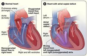

The septum is the wall that separates the right and left sides of the heart. A hole in the wall between the two upper chambers is called an atrial septal defect, or ASD. This is one of the least complex forms of congenital heart defect, and was one of the first types to be repaired surgically. Normally, low-oxygen blood entering the right side of the heart stays on the right side, and oxygen-rich blood stays on the left side of the heart, where it is then pumped to the body. When a defect or "hole" is present between the atria (or upper chambers), some oxygen-rich blood leaks back to the right side of the heart. It then goes back to the lungs even though it is already rich in oxygen. Because of this, there is a significant increase in the blood that goes to the lungs.
There are three different kinds of ASDs. The most common form of ASD is the secundum defect which usually occurs as an isolated defect. The primum ASD is associated with a cleft in the mitral valve which may also causing the valve to leak. The third kind of ASD is the sinus venosus defect, located in the superior portion of the atrial septum and typically associated with abnormal drainage of the right upper pulmonary vein.
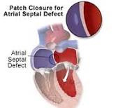
Atrial septal defects can vary greatly in size. Some ASDs will close on their own and no surgery is needed. Some ASDs are closed in the catheterization lab and do not require open-heart surgery. Certain devices such as the Amplatzer Occluder, the CardioSEAL, Helex, and Clamshell Device are currently being used or have been used in the past. Some ASDs will need to be corrected with open heart surgery to restore normal blood circulation and/or to repair subsequent damage which has occurred in the heart. Many ASDs are not detected until adulthood. Left untreated for decades, potential problems include lung disease, exercise intolerance, heart rhythm abnormalities, shortened life expectancy and the increased risk of a stroke.
Atrial septal defects occur in 5 to 10 percent of all children born with congenital heart disease. For unknown reasons, girls have atrial septal defects twice as often as boys.
There are three different kinds of ASDs. The most common form of ASD is the secundum defect which usually occurs as an isolated defect. The primum ASD is associated with a cleft in the mitral valve which may also causing the valve to leak. The third kind of ASD is the sinus venosus defect, located in the superior portion of the atrial septum and typically associated with abnormal drainage of the right upper pulmonary vein.
Atrial septal defects can vary greatly in size. Some ASDs will close on their own and no surgery is needed. Some ASDs are closed in the catheterization lab and do not require open-heart surgery. Certain devices such as the Amplatzer Occluder, the CardioSEAL, Helex, and Clamshell Device are currently being used or have been used in the past. Some ASDs will need to be corrected with open heart surgery to restore normal blood circulation and/or to repair subsequent damage which has occurred in the heart. Many ASDs are not detected until adulthood. Left untreated for decades, potential problems include lung disease, exercise intolerance, heart rhythm abnormalities, shortened life expectancy and the increased risk of a stroke.
Atrial septal defects occur in 5 to 10 percent of all children born with congenital heart disease. For unknown reasons, girls have atrial septal defects twice as often as boys.
