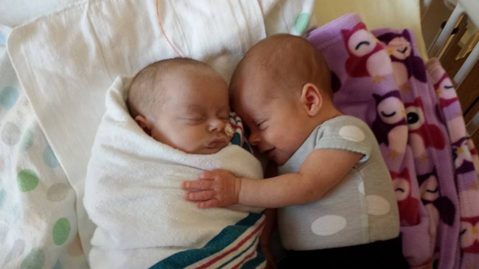
Terra and her twin sister Luna were born on February 9, 2014 at 35 weeks gestation. The journey of these two miracles actually began many months before this. Kaylin found out when she was 7 weeks pregnant that she was expecting twins - not only twins but Mo Mo Twins. Most of you have probably never heard of Mo Mo twins, I know that I had not until she was diagnosed. I had always thought that there were only two types of twins, identical and faternal. Well guess what? I was mistaken. There are actually four different types of identical twins depending upon when the egg splits. Our twins are Mo Mo which means that they were very high risk with Momma Kaylin having to be monitored very closely with mutiple ultrasounds and more than two months of being in the hospital on bed rest to get them here safely. Upon seeing the Maternal Fetal Medicine physican she was told that IF she made it to 24 weeks she would be put in the hospital on bedrest but that there was not much they could do before that point to save them if their cords became entangled. So it was just a wait and pray time. At her 20 week ultrasound Terra was diagnosed with multiple heart defects so this complicated the pregnancy even more. Normally the doctors do not allow Mo Mo babies to go past 32 to 34 weeks gestation because of the high risk involved but with Terra's heart defects they needed her to stay in the womb as long as possible to get her to a big enough size that she could be operated on. Terra and Luna also have an older brother and sister so Kaylin had to be away from them during her inpatient stay and only be able to see them on weekends.
Mo Mo Twins:
http://en.wikipedia.org/wiki/Monoamniotic_twins
Monoamniotic twins are identical twins that share the same amniotic sac within their mother’s uterus.Monoamniotic twins are always identical, and always monochorionic as well (sharing the same placenta), and are sometimes termed Monoamniotic-Monochorionic ("MoMo") twins.They also share the placenta, but have two separate umbilical cords. Monoamniotic twins develop when an embryo does not split until after formation of the amniotic sac, at about 9 days after fertilization.
Monoamniotic twins are rare, with an occurrence of 1 in 60,000 pregnancies, corresponding to about 1% of twin pregnancies.
The survival rate for monoamniotic twins has been shown to be as high as 81%to 95%in 2009 with aggressive fetal monitoring, although previously reported as being between 50%to 60%. Causes of mortality and morbidity include:
Cord entanglement: The close proximity and absence of amniotic membrane separating the two umbilical cords makes it particularly easy for the twins to become entangled in each other’s cords, hindering fetal movement and development. Additionally, entanglement may cause one twin to become stuck in the birth canal during labor and expulsion. Cord entanglement happens to some degree in almost every monoamniotic pregnancy.
Cord compression: One twin may compress the other’s umbilical cord, potentially stopping the flow of nutrients and blood and resulting in fetal death.
Twin-to-twin transfusion syndrome (TTTS): One twin receives the majority of the nourishment, causing the other twin to become undernourished. TTTS is much more difficult to diagnose in monoamniotic twins than diamniotic ones, since the standard method otherwise is to compare the fluid in the sacs. Rather, TTTS diagnosis in monoamniotic twins relies on comparing the physical development of the twins.
Regular and aggressive fetal monitoring is recommended for cases of monoamniotic twins to look for cord entanglement beginning after viability. Many women enter inpatient care, with continuous monitoring, preferably in the care of a perinatologist, an obstetrician that specialises in high risk pregnancies.
All monoamniotic twins are delivered prematurely by cesarean section, since the risk of cord entanglement and/or cord compression becomes too great in the third trimester. The cesarean is usually performed at 32, 34 or 36 weeks. Many monoamniotic twins experience life-threatening complications as early as 26 weeks, motivating immediate delivery. However, delivery around 26 weeks is associated with life-threatening complications of preterm birth. Steroids may be administered to stimulate the babies' lung development and decrease the risk of infant respiratory distress syndrome. Natural birth rather than cesarean section causes cord prolapse, with the first baby delivered pulling the placenta shared with the baby being left inside.
Terra's defects:
This article will help to explain.
http://www.cincinnatichildrens.org/health/t/tricuspid/
Tricuspid atresia is a type of congenital heart disease in which the valve between the right atrium and right ventricle fails to develop.
Blood that returns from the body to the right atrium cannot directly enter the right ventricle, and must pass through a hole in the atrial septum (atrial septal defect) into the left atrium and then the left ventricle.
There are several anatomic variations that influence the symptoms and course of treatment in any given patient.
There may be a hole in the ventricular septum, called a ventricular septal defect (VSD).
The aorta and pulmonary artery may be normally positioned and aligned with the appropriate ventricle or they may be reversed, a condition called transposition of the great arteries.
If the ventricular septal defect is small or absent, and the great arteries are normally positioned, blood flows from the left ventricle out the aorta to the body. In this situation very little, if any, blood can get to the lungs resulting in very low oxygen levels in the infant.
In a newborn, blood can reach the lungs to pick up oxygen as long as a connection between the aorta and pulmonary artery called the ductus arteriosus remains open. The “ductus” is an important vessel while the baby is still in the womb because it allows the blood from the baby's heart to return to the placenta, which does the job of the lungs before birth.
This vessel is exquisitely sensitive to oxygen, so when the baby is born, it typically narrows down (or closes completely) after 24 to 48 hours in response to the oxygen levels in the air breathed by the child. An intravenous medication called prostaglandin (PGE) can keep this important vessel open after birth.
If a ventricular septal defect is present and the great arteries are in their normally related position, blood from the left ventricle can reach the lungs through the ventricular septal defect. This channel is often very narrow, and the right ventricle very underdeveloped, so a less than normal amount of blood goes to the lungs.
Finally, if there is transposition of the great arteries, blood reaches the lungs easily because the pulmonary artery is directly connected to the left ventricle. But blood can only reach the body through the ductus arteriosus or the ventricular septal defect if there is one.
Treatment of Terra's Defects:
Note: A newborn baby's heart is the size of a walnut.
The diagnosis of tricuspid atresia with too little blood flow to the lungs or to the body requires immediate medical treatment. In a newborn (less than 1 to 2 weeks old), a medication called PGE can be given intravenously to reopen the connection (ductus arteriosus) between the pulmonary artery and aorta and improve blood flow to the lungs or body.
Children with tricuspid atresia and too little pulmonary blood flow will require surgery to establish a connection between the arteries to the body and the arteries to the lungs. This type of operation is call a modified Blalock-Taussig (BT) shunt, and involves the placement of a small Gore-Tex tube between the artery to the arm (subclavian artery) and the arteries to the lungs (pulmonary artery).
If the problem is too much pulmonary blood flow (tricuspid atresia with a large ventricular septal defect), blood flow to the lungs will usually need to be limited to protect the lungs from becoming damaged by too much blood flow. This can be accomplished by placing a band around the pulmonary artery so that blood flow to the lungs is limited in a controlled way.
Finally, if the problem is inadequate blood flow through the aorta (tricuspid atresia with ventricular septal defect and transposition of the great arteries), blood from the normal size left ventricle will need to be routed to the aorta, and the aorta will usually need to be reconstructed. Pulmonary blood flow can then be established by placement of a modified BT shunt. This is essentially the Norwood procedure used for hypoplastic left heart syndrome.
Whatever operation is necessary in the newborn period, children can expect to undergo further heart surgery by the age of 3 to 6 months. This is true whether the child has too much or too little pulmonary or systemic blood flow in the newborn period and, therefore, did not require any surgery at that time.
The operation at 3 to 6 months is called a bidirectional Glenn. The superior vena cava is detached from the heart and connected directly to the pulmonary artery, and the BT shunt is removed. This allows blood from the upper body to flow directly to the lungs to pick up oxygen without having to be pumped by the heart. It also prevents blood that already has oxygen from returning to the lungs, and, thereby, keeps the heart from doing unnecessary work. After this operation, however, there is still blood returning from the body through the inferior vena cava going directly back to the body without first passing through the lungs. Because of this, some level of cyanosis will persist.
Between the ages of 2 and 5 years, children with tricuspid atresia will be ready for the third operation required to optimize their circulation. This operation is called the Fontan procedure, and involves connection of the inferior vena cava directly to the pulmonary artery, which forces all blood returning from the body to pass through the lungs and pick up oxygen before being pumped to the body. This allows a more normal color in the skin and lips as well due to a more normal oxygen saturation in the blood.
